UNIT 1
Coronary Artery Disease & Acute Coronary Syndrome
Covers: Lewis's Ch. 37 (pp. 819–859) · Lecture 1, Part 1
1.1 Coronary Anatomy — Why the Map Matters
Every question about infarct location traces back to one fact: each coronary artery feeds a specific region of myocardium. When an artery occludes, the muscle downstream of the blockage becomes ischemic, then necrotic. Learn which artery feeds which wall now and the 12-lead section later becomes recognition instead of memorization.
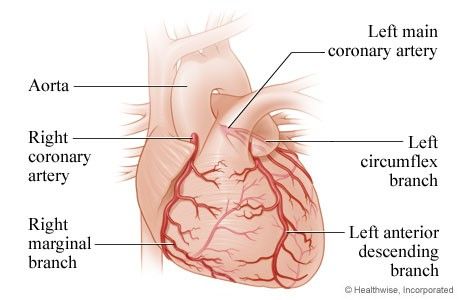
- Left anterior descending (LAD) — anterior wall and interventricular septum. The largest territory, sometimes called the widow-maker.
- Left circumflex — lateral wall of the left ventricle.
- Right coronary artery (RCA) — inferior wall. (Background worth carrying, though it is not on her slides: the RCA also feeds the SA and AV nodes in most people, which is why inferior MI so often arrives with bradycardia and AV block.)
1.2 Coronary Artery Disease and Atherosclerosis
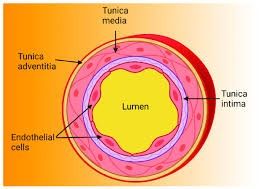
CAD is the most common cardiovascular disease and the leading cause of death in the United States. It sits in the category of atherosclerosis — lipid deposits forming within the tunica intima, the innermost layer of the artery wall.
The step that starts everything is endothelial injury. A layer of endothelial cells normally sits between the intima and the blood, keeping substances out of the wall that do not belong there. Damage that barrier and lipoproteins can carry cholesterol from the blood into the intima, where plaque begins.
The three stages of CAD
| Stage | What is happening | Clinical meaning |
|---|---|---|
| 1. Fatty streak | Fatty streaks appear on smooth muscle cells. Present in many people by about age 20. | Reversible window. Lipid-lowering strategies — diet, exercise, smoking cessation, statins if indicated — slow progression. |
| 2. Fibrous plaque | Collagen covers the fatty streak. The lumen narrows and blood flow to distal tissue falls. | Where chronic stable angina lives — a fixed narrowing that cannot meet increased demand. |
| 3. Complicated lesion | Plaque grows, inflammation draws platelets, thrombus forms and adheres to the wall. Plaque may rupture. | The last and most dangerous stage. This is the substrate for acute coronary syndrome. |
1.3 Risk Factors
| Modifiable | Nonmodifiable |
|---|---|
| BP > 120/80 | Increasing age |
| Diabetes | Ethnicity |
| Total cholesterol > 200 mg/dL | Gender |
| Triglycerides ≥ 150 mg/dL | Genetic predisposition |
| LDL > 130 mg/dL | Family history of heart disease |
| HDL < 40 (men) or < 50 (women) mg/dL | |
| Metabolic syndrome | |
| Physical inactivity | |
| Tobacco use / substance abuse | |
| Psychosocial risk factors |
Metabolic syndrome
Also called syndrome X. A cluster of conditions occurring together that raises the risk of heart disease, stroke, and type 2 diabetes: increased blood pressure, high blood sugar, excess body fat around the waist, and abnormal cholesterol and triglyceride levels.
1.4 Collateral Circulation
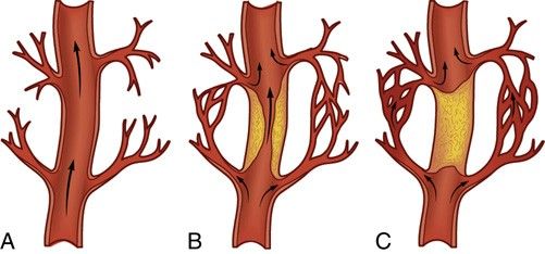
Named explicitly on the posted exam study guide. Collateral circulation is arterial anastomoses — new connections that develop around a blockage so blood has an alternate path to the tissue beyond it. The instructor's framing: think of it as a detour.
1.5 Nursing Management of CAD
- Identify patients at high risk — take an adequate health history covering family history, diabetes, hypertension, and smoking.
- Assess educational background and health literacy so teaching lands. Do not use dense medical terminology with a patient who cannot follow it.
- Educate on managing modifiable risk factors and developing health-promoting behaviors.
- Medication compliance and education.
- Help the patient set realistic goals. The instructor calls this one of the most important things — telling someone to exercise six or seven days a week and overhaul their diet at once is overwhelming and does not stick. Smaller, reachable goals produce consistency.
1.6 The ACS Taxonomy — Hold This Structure
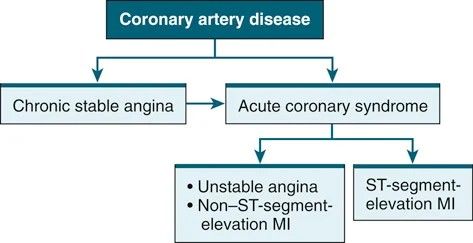
Almost every ACS question depends on knowing where a presentation sits in this hierarchy. CAD is the umbrella term. Underneath it, one branch is chronic stable angina; the other is acute coronary syndrome, which contains unstable angina, NSTEMI, and STEMI.
1.7 Chronic Stable Angina
Myocardial demand for oxygen exceeds the ability of the coronary arteries to supply it. The result is myocardial ischemia, which the patient experiences as chest pain. The narrowing is fixed, so the pain is predictable — the same character, onset, and duration each time, brought on by the same triggers and relieved when the trigger is removed.
How patients actually describe it
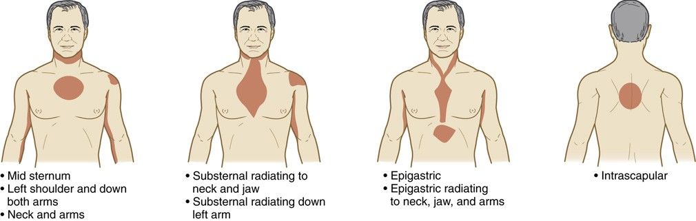
- Often not called chest pain at all — pressure, heaviness, discomfort, squeezing, tightness, a suffocating sensation, or something sitting on the chest.
- Dyspnea and fatigue.
- No change with position or breathing — this is what separates it from pleuritic or musculoskeletal pain.
- Usually substernal, radiating to neck, jaw, shoulders, or arms; may present between the shoulder blades (interscapular).
- May be described as indigestion or epigastric burning.
- Lasts a few minutes and resolves once the aggravating factor is gone.
- Assess it with LOUDCARTS, the mnemonic on her slide.
Precipitating factors
| Trigger | Why it provokes angina |
|---|---|
| Waking or early morning | Circadian surge in sympathetic tone. |
| A large meal | Blood is redirected to the GI tract for digestion, so less reaches the coronary arteries. The instructor's example: emergency departments fill the day after a holiday. |
| Physical exertion | Raises myocardial oxygen demand directly. |
| Sexual activity | Same demand mechanism as exertion. |
| Stimulants | ↑ HR, ↑ BP, and ↑ myocardial oxygen demand, plus vasoconstriction that lowers supply — demand up and supply down at once. |
| Strong emotions, extreme temperatures | Sympathetic activation. |
| Tobacco or environmental smoke | Vasoconstriction reduces oxygen and blood supply to the myocardium. |
EKG in chronic stable angina
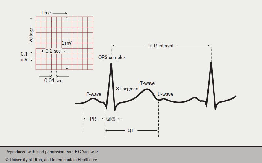
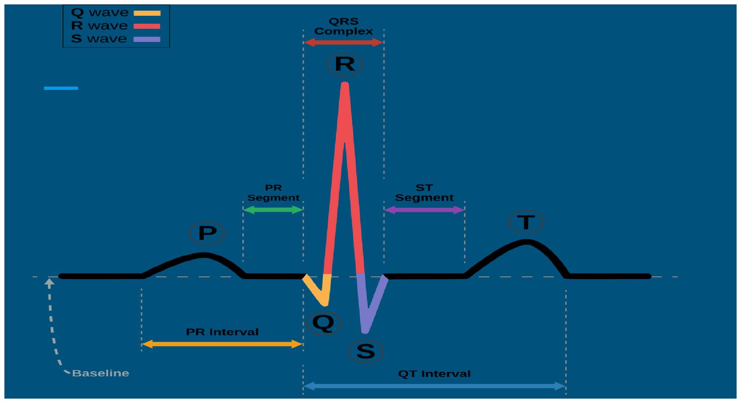
ST segment depression and/or T wave inversion — the signature of ischemia without infarction. It resolves once oxygen supply is restored. Goals of treatment are simple and worth memorizing as a pair: reduce angina symptoms, and reduce the risk of MI and death.
1.8 Silent Ischemia
- Ischemia occurring without subjective symptoms.
- Seen in patients with diabetes mellitus, because neuropathy affects the nerves innervating the cardiovascular system. More likely in advanced diabetes where neuropathy has developed as a complication.
- EKG changes still appear — ST depression and/or T wave inversion. The ischemia is silent to the patient, not to the monitor.
1.9 Prinzmetal's Angina
A rare form occurring at rest, with no physical exertion, caused by hyperactivity of vascular smooth muscle producing spasm of the vessel. It can occur with or without CAD.
- Risk factors: history of migraines, Raynaud's phenomenon, heavy smoking.
- Substances and drugs: alcohol, cocaine, sumatriptan (which narrows blood vessels — hence the migraine link).
- EKG: transient ST segment elevation — it appears and then goes away.
- Patients may report short bursts of chest pain at the same time each day.
- Treatment: calcium channel blockers and sublingual nitroglycerin; remove the offending agent. Pain may also resolve spontaneously when the spasm stops.
- Long-acting nitrates are also used for Prinzmetal's.
1.10 Unstable Angina
A previously stable chronic plaque becomes unstable. It ruptures, releasing its lipid core into the vessel; platelets aggregate and a thrombus forms, partially or completely occluding the vessel. Partial occlusion produces unstable angina or NSTEMI; complete occlusion produces STEMI.
- Pain is new in onset and occurs at rest, not only with activity.
- In a patient with known chronic stable angina, the warning sign is a change in their normal pattern — increasing frequency, increasing duration, or a different character. They know their usual pain; when they say this one is different, listen.
- Lasts 10 minutes or more.
- EKG: ST depression and/or T wave inversion.
- Needs to be treated immediately.
1.11 NSTEMI and Cardiac Biomarkers
A myocardial infarction is the abrupt stoppage of blood flow in a vessel caused by platelet aggregation, blocking blood and oxygen to the heart. Everything downstream of the blockage becomes necrotic — irreversible myocardial cell death. In NSTEMI the occlusion is not complete, so the EKG may or may not show ST depression or T wave inversion in the leads facing the infarct.
| Biomarker | Rises | Peaks | Returns to normal | Notes |
|---|---|---|---|---|
| Troponin T (cTnT) and Troponin I (cTnI) | 4–6 hr after MI onset | 10–24 hr | 10–14 days | Cardiac specific. The standard. |
| High-sensitivity troponin (hs-cTn) | Within 1 hr of injury | — | Stays high 7–14 days | Faster diagnosis. Second level drawn at 2–3 hr. |
| CK-MB | 6 hr | 18 hr | 24–36 hr | Less sensitive than troponin; helps quantify damage. |
1.12 STEMI
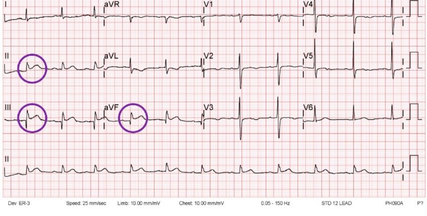
An occlusive thrombus completely blocks the vessel, producing ST elevation in the leads facing the infarcted area. This is an emergency.
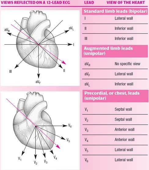
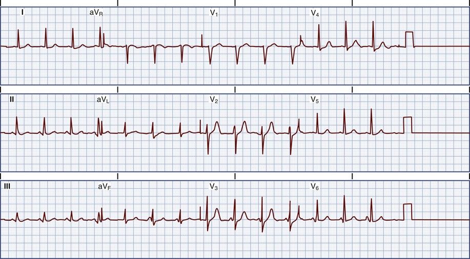
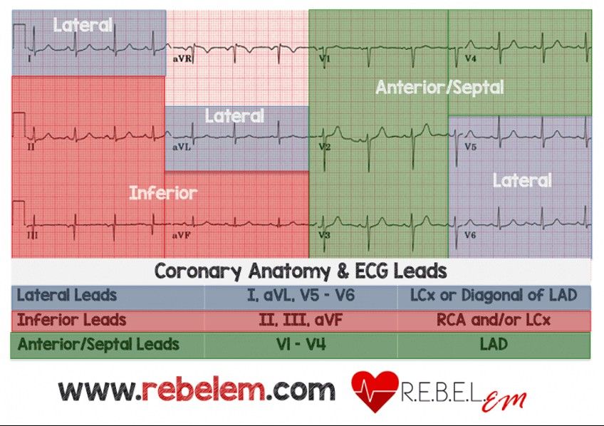
Contiguous leads means two leads looking at the same region of the heart. For an inferior STEMI, the elevation must appear in two of II, III, and aVF.
1.13 Locating the Infarct
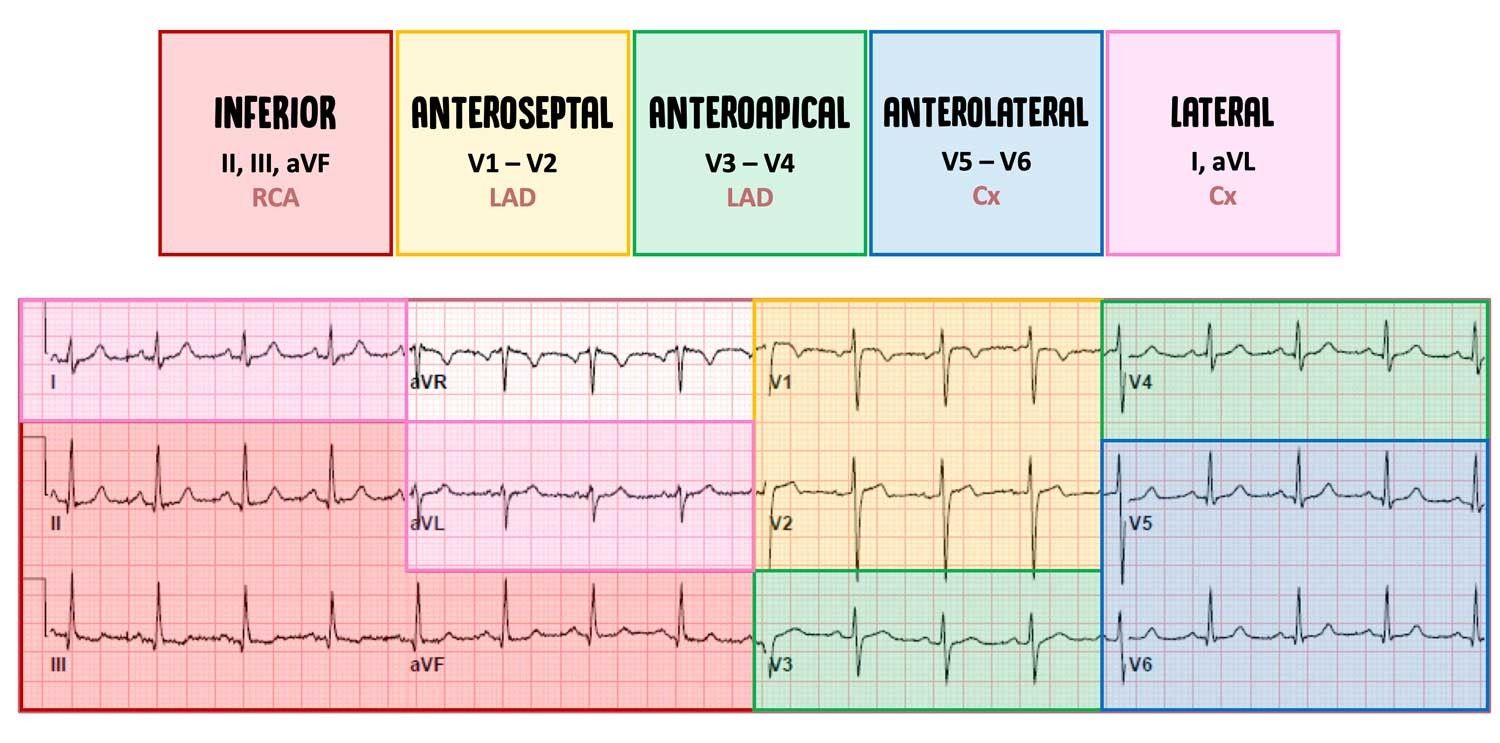
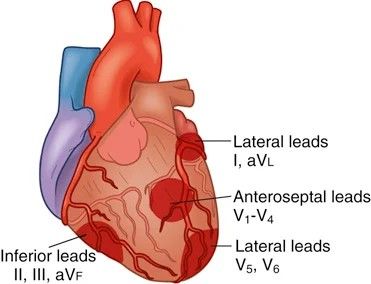
Named on the posted exam study guide as "location of myocardial infarction." The leads showing elevation tell you which wall is infarcting, which tells you which artery is occluded.
| Wall | Leads facing it | Reciprocal leads | Artery |
|---|---|---|---|
| Septal | V1, V2 | II, III, aVF | Left anterior descending |
| Anterior | V3, V4 | II, III, aVF | Left anterior descending |
| Lateral, low | V5, V6 | II, III, aVF | LAD or circumflex |
| Lateral, high | I, aVL | II, III, aVF | Circumflex |
| Inferior | II, III, aVF | I, aVL, V5, V6 | Right coronary artery |
1.14 Percutaneous Coronary Intervention
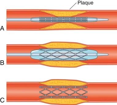
First-line treatment for confirmed STEMI, with the goal of opening the blocked artery within 90 minutes of presentation at a PCI-capable hospital. Cardiac catheterization assesses the location and extent of the blockage; a guidewire is advanced from the femoral or radial artery to the coronary vessel, a balloon is inflated to push the plaque aside, and in most cases a stent is placed to hold the artery open.
Antiplatelet and anticoagulation after PCI
- Stents are thrombogenic — clots form on them.
- During PCI: unfractionated heparin or low molecular weight heparin (enoxaparin / Lovenox).
- After PCI: dual antiplatelet therapy (DAPT) — aspirin plus clopidogrel (Plavix).
- Aspirin for life. Clopidogrel for 12 months — or only 3–6 months with newer-generation drug-eluting stents.
- DAPT continues until the intimal lining grows over the metal stent, restoring a smooth surface for blood flow.
Benefits and complications
| Benefits | Potential complications |
|---|---|
| Faster reperfusion | Dissection or rupture of the coronary artery |
| Quicker and easier than surgery | Abrupt artery closure |
| Faster recovery | Acute stent thrombosis causing acute MI |
| Stroke, from dislodged plaque traveling to cerebral arteries | |
| Bleeding and infection | |
| Failure to cross the blockage with balloon angioplasty — this patient may need CABG instead | |
| Dysrhythmias, most commonly atrial fibrillation |
Nursing care after PCI
Named specifically on the posted exam study guide.
- Monitor vital signs and compare against baseline.
- Listen to heart and breath sounds.
- Assess neurovascular status of the affected extremity every 15 minutes for the first hour, then per agency policy — that is four checks in the first hour. Check distal to the site: pedal and posterior tibial pulses, capillary refill, temperature, color.
- Assess the catheter insertion site for hematoma, bleeding, and bruit.
- Place a compression device over the arterial site to achieve hemostasis, per agency policy.
- Monitor the ECG for dysrhythmias.
- Monitor for chest pain. Pain should be relieved after PCI. If it is not, the patient may have another blockage or the PCI may have failed — report to the provider right away.
- Teach the patient and caregiver about discharge medications.
1.15 Thrombolytic Therapy
Indicated when the patient is at a facility without PCI capability. These drugs break up the fibrin meshwork in the clot, dissolving the occlusive thrombus and restoring perfusion.
The instructor answered a class question about these drugs: yes, thrombolytics from this same group are what the ED uses for ischemic stroke — never hemorrhagic, since that patient is already bleeding. (The specific agent she named is garbled in the recording, so it is left unnamed here rather than guessed at; alteplase and tenecteplase are both used for stroke.)
Absolute contraindications
She read these aloud from the textbook chart. The common thread is simple — anything that means the patient is bleeding, has bled into the head, or is about to.
- Active internal bleeding
- History of intracranial hemorrhage
- Intracranial or intraspinal surgery within 2 months
- Known structural or vascular abnormality (e.g. arteriovenous malformation)
- Known intracranial cancer, primary or metastatic
- Ischemic stroke within the past 3 months
- Severe uncontrolled hypertension
- Significant closed-head or facial trauma within the past 3 months
- Suspected aortic dissection
Nursing points during and after
- Place 2–3 IV lines and draw baseline labs before starting; complete all other invasive procedures first, to reduce bleeding risk.
- The most reliable sign of reperfusion is return of the ST segment to baseline. Others: resolution of chest pain, and an early rapid rise in cardiac biomarkers as the necrotic cells wash out.
- Reperfusion dysrhythmias are common and generally self-limiting.
- Main complication is bleeding. Assess neurologic status regularly for signs of cerebral bleeding.
- Reocclusion is a major concern — IV heparin is started, and the patient should be moved to a PCI-capable facility.
1.16 Coronary Artery Bypass Graft
The posted exam study guide asks two things about CABG: when it is indicated, and nursing care after surgery.
The procedure
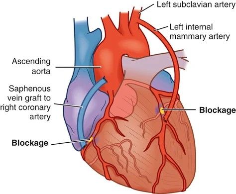
- Sternotomy — the chest is opened down the middle and the ribs moved to reach the heart.
- Cardiopulmonary bypass — the heart is stopped and a machine oxygenates and circulates the blood, doing the work of the heart and lungs.
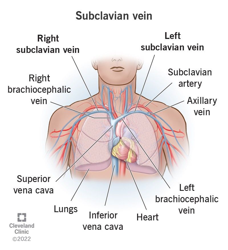
- Most common conduit: the left internal mammary artery (LIMA), which comes off the subclavian artery and is closest to the heart.
- Alternatives: saphenous vein from the leg, or radial artery from the forearm.
- Off-pump CABG — still a sternotomy, but performed on a beating heart using mechanical stabilizers, so no bypass machine is needed.
Post-operative care
- ICU stay for 24–48 hours.
- Most patients extubated within about 6 hours.
- Transferred to step-down in 24–48 hours if stable.
- Monitor hemodynamics — BP, HR, and the invasive lines.
- Expect a full inventory of lines: arterial line for continuous BP, pleural and/or mediastinal chest tubes, continuous ECG, endotracheal tube, epicardial pacing wires, Foley catheter, possible NG tube for gastric decompression.
Complications — mostly from the bypass machine
| Complication | Why it happens |
|---|---|
| Systemic inflammation | Tissue manipulation during surgery plus exposure of blood to the artificial surfaces of the bypass circuit. |
| Bleeding and anemia | Surgical blood loss and circuit-related coagulopathy. |
| Fluid and electrolyte imbalances | Magnesium and potassium commonly need replacement. |
| Infection | Watch for fever, elevated heart rate, low blood pressure, and incision drainage, redness, warmth, swelling, or increasing pain. |
| Hypothermia | Cooling during bypass; use warming blankets. |
| Atrial fibrillation | Occurs in 20–50% of patients — the single most quotable number in this section. |
Nursing management after CABG
- Assess for bleeding from the chest tube and incision site.
- Monitor hemodynamics and check fluid status.
- Replace blood and electrolytes as ordered.
- Restore temperature with warming blankets.
- Wound care to the incision site.
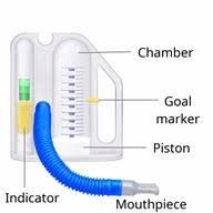
- Manage pain — the chest was cut open, and uncontrolled pain prevents deep breathing.
- Prevent VTE — sequential compression devices and early ambulation.
- Prevent respiratory compromise — incentive spirometry, deep breathing, and coughing to expand the alveoli and prevent atelectasis and pneumonia.
1.17 The Patient Arriving With Chest Pain
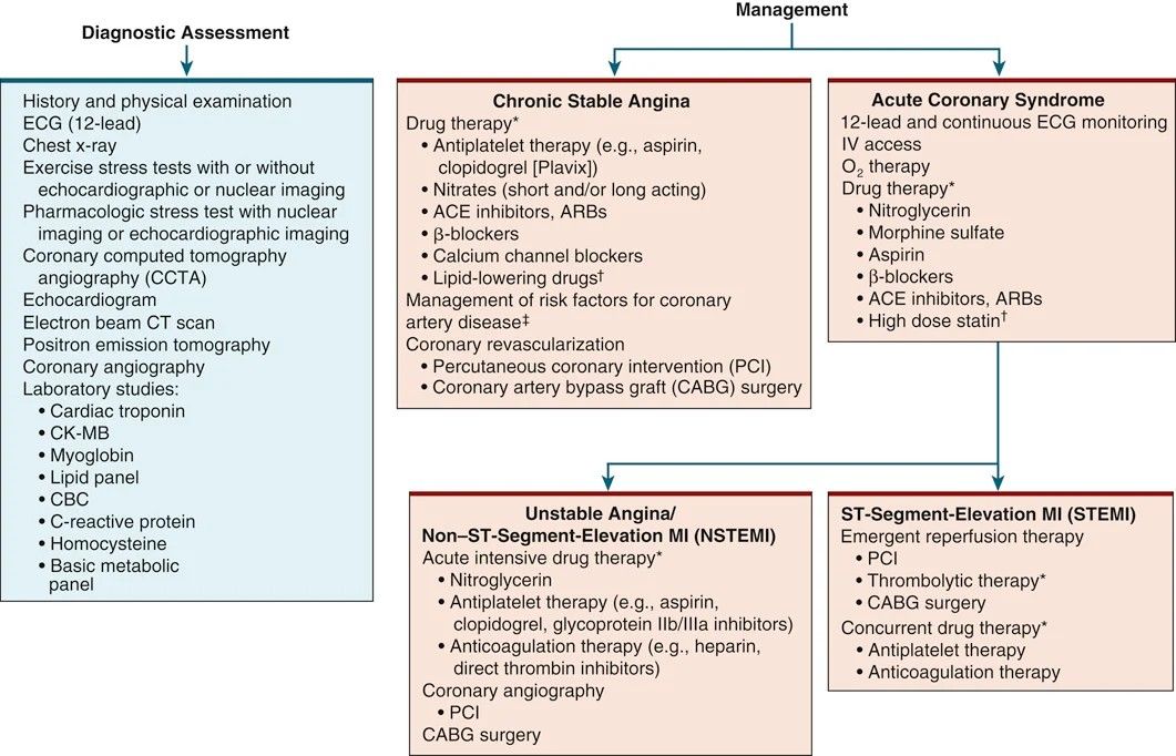
A high-yield sequence, because it is naturally written as a "which action would the nurse take first" question.
1.18 Drug Therapy for Angina and MI
Nitrates
| Short-acting | Long-acting | |
|---|---|---|
| Examples | Sublingual nitroglycerin tablets, translingual spray | Isosorbide dinitrate (Isordil), isosorbide mononitrate (Imdur) |
| Use | Acute episode of angina | Reduce the frequency of angina attacks; treat Prinzmetal's |
| Action | Dilate peripheral vessels (↓ SVR, ↓ preload) and dilate coronary arteries and collateral vessels | Same mechanism, sustained |
| Key teaching | Relief in 5 min, lasts 30–40 min. If unchanged or worse at 5 min → call 911. If relieved, may repeat every 5 min for a total of 3 doses. | Tolerance develops → 10–14 hour nitrate-free period. Short-acting nitrates can still be used for breakthrough pain. |
| Side effects | Hypotension, dizziness, headache, flushing. Change positions slowly. | Headache is the main one. |
The other drug classes
| Class | Examples | What it does | Why it helps |
|---|---|---|---|
| ACE inhibitors | "-pril" — lisinopril, captopril | Vasodilation, reduced blood volume | Prevent or reverse ventricular remodeling after MI |
| ARBs | "-sartan" — losartan, candesartan | Same effect via a different receptor | Used when ACE inhibitors are not tolerated (cough, angioedema) |
| Beta blockers | "-lol" — metoprolol, labetalol, propranolol | ↓ contractility, ↓ HR, ↓ SVR, ↓ BP | ↓ myocardial oxygen demand, resolving angina |
| CCB — dihydropyridines | amlodipine, nifedipine | More vasodilation | Mainly blood pressure control |
| CCB — nondihydropyridines | verapamil, diltiazem | ↓ contractility and ↓ HR | Rate control |
How calcium channel blockers work, from first principles. Calcium normally causes vasoconstriction of arterial smooth muscle and drives cardiac contraction — whenever calcium enters, muscle excites, contracts, and constricts. Blocking calcium entry relaxes vascular smooth muscle and reduces contractility. The net effect is decreased heart rate, decreased contractility, and coronary vasodilation, all lowering myocardial oxygen demand.
Lipid-lowering drugs
| Drug class | Mechanism | Effect |
|---|---|---|
| Statins — rosuvastatin (Crestor), simvastatin (Zocor) | Inhibit cholesterol synthesis in the liver and increase hepatic LDL receptors. Best tolerated. | ↓ LDL. Side effects: rhabdomyolysis, liver damage, myalgia. |
| Niacin | Inhibits synthesis and secretion of VLDL and LDL | ↓ LDL, ↓ triglycerides, ↑ HDL |
| Fibric acid derivatives — fenofibrate (Tricor), gemfibrozil (Lopid) | ↓ hepatic synthesis and secretion of VLDL | ↓ VLDL, ↓ triglycerides, ↓ LDL, ↑ HDL |
| ATP-citrate lyase inhibitor — bempedoic acid (Nexletol) | Inhibits synthesis and secretion of VLDL and LDL | ↓ LDL, ↓ triglycerides, ↑ HDL |
| Bile-acid sequestrants — colesevelam (Welchol), colestipol, cholestyramine | Bind bile acids in the intestine into an insoluble complex excreted in feces | ↓ LDL and cholesterol |
| Cholesterol absorption inhibitor — ezetimibe (Zetia) | Inhibits intestinal absorption of cholesterol | ↓ LDL, ↑ HDL. Often added to a statin. |
Anticoagulants and antiplatelets
The distinction the instructor draws: anticoagulants prevent clots from forming; antiplatelets prevent platelets from aggregating onto a clot that already exists.
| Anticoagulants | Antiplatelets |
|---|---|
| Low molecular weight heparin: enoxaparin (Lovenox) | Aspirin |
| Unfractionated heparin | Clopidogrel (Plavix) |
| Vitamin K antagonist: warfarin (Coumadin) | Ticagrelor (Brilinta) |
| Factor Xa inhibitors: apixaban (Eliquis), rivaroxaban (Xarelto), fondaparinux (Arixtra) | Prasugrel (Effient) |
| Direct thrombin inhibitors: bivalirudin (Angiomax), argatroban | Glycoprotein IIb/IIIa inhibitors: abciximab (ReoPro), eptifibatide (Integrilin), tirofiban (Aggrastat) |
Warfarin requires frequent INR monitoring, which is why many providers now favor apixaban or rivaroxaban. Glycoprotein IIb/IIIa inhibitors work by stopping fibrinogen from binding and forming bridges between platelets.
Beyond drugs
- Blood pressure control · smoking cessation · cardiac rehab
- Diet · exercise · diabetes management
- Depression management · patient education · flu vaccination
1.19 Sudden Cardiac Death
Abrupt, unexpected death resulting from a variety of cardiac causes. The posted exam study guide asks specifically about common causes.
A patient who survives SCD remains at risk of another event, because scarred myocardium creates ongoing electrical instability.
Interprofessional and nursing care after SCD
- Work the patient up to determine whether an MI caused the event.
- Cardiac catheterization, PCI, or CABG as indicated.
- Holter monitor — worn on the outside of the skin, typically for 24 hours, sometimes one to two weeks — or an implantable loop recorder.
- Electrophysiology study (EPS).
- Drug therapy.
- Teach family and caregivers CPR and AED use.
- Implantable cardioverter-defibrillator (ICD) — the most common approach to improving survival from SCD and preventing recurrence. Covered in full in Unit 2.